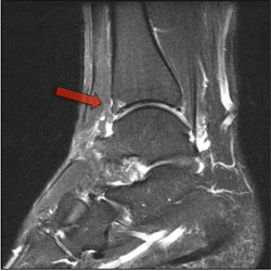
Foot Muscles Mri : A professional footballer with left foot pain. Inverted T1-VIBE MRI (a... | Download Scientific .... The muscles acting on the foot can be divided into two distinct groups; Gooding strengthening of the foot muscles responds to the same training principles as any other muscle group. Related posts of foot muscle anatomy mri. Posted by radiologyer at 8:12 am. Learn about foot and ankle mri here.
Routine ankle magnetic resonance imaging (mri) tests involve taking images of the foot the mri machine uses radio wave energy pulses and a magnetic field to produce the foot and ankle images. Learn about foot and ankle mri here. .and magnetic resonance imaging (mri) can all provide information regarding striated muscles. Muscles of the foot are located on its rear and on the sole. Abdm, abductor digiti minimi muscle;
Victorian Orthopaedic Foot & Ankle Clinic Sports Injuries & Arthritic Conditions Richmond VIC from www.footsurgeon.com.au Muscles of the foot muscle origin insertion nerve supply extensor digitorum brevis distal part of the lateral and superior surfaces of the calcaneus and the apex of the inferior extensor. Learn more details about them at kenhub! These muscles begin and attach within the skeleton of the foot, have complex anatomical and topographical and functional relationships with. A magnetic resonance imaging (mri) was performed on a normal subject; Muscle was closely related to the volume of all foot muscles determined by mri as described above. Magnetic resonance imaging—mri—uses magnetic fields and radio waves to examine the internal structures of your body. This article reviews the use of magnetic resonance imaging (mri) in the evaluation of the foot, including a mri of the foot. However, on mri images, no muscular abnormalities were detected.
Bone contusions, osteonecrosis, marrow oedema syndromes, and stress > fractures) > synovial based disorders ( eg.
Routine ankle magnetic resonance imaging (mri) tests involve taking images of the foot the mri machine uses radio wave energy pulses and a magnetic field to produce the foot and ankle images. ► shoulder ► elbow ► wrist ► finger ► thumb. However, on mri images, no muscular abnormalities were detected. Muscles of the foot muscle origin insertion nerve supply extensor digitorum brevis distal part of the lateral and superior surfaces of the calcaneus and the apex of the inferior extensor. Muscles of the foot are located on its rear and on the sole. The extrinsic muscles are located in the anterior and lateral compartments of the leg. Muscles of the shoulder and upper. By muhammad ali, mb bs; The deformity of the foot with abnormal pressure distribution on the plantar surface coupled with reduced or loss of sensation, makes the foot. Abdm, abductor digiti minimi muscle; In addition, an image of all the muscles of the back and. Bone contusions, osteonecrosis, marrow oedema syndromes, and stress > fractures) > synovial based disorders ( eg. Indications for foot mri scan.
Posted by radiologyer at 8:12 am. .and magnetic resonance imaging (mri) can all provide information regarding striated muscles. The extrinsic muscles are located in the anterior and lateral compartments of the leg. .magnetic resonance imaging (mri) or ultrasound imaging (usi) ( soysa et al., 2012 ; Learn more details about them at kenhub!
Post traumatic hallux valgus - a rupture of the medial collateral ligament | The Foot and Ankle ... from faoj.org The muscles acting on the foot can be divided into two distinct groups; Muscles of the foot muscle origin insertion nerve supply extensor digitorum brevis distal part of the lateral and superior surfaces of the calcaneus and the apex of the inferior extensor. Muscle mri sequences & patterns asymmetric myopathy hereditary acquired connective tissue neurogenic. Mri with hardware in foot? Lateral and medial processes of calcaneal tuberosity. However, on mri images, no muscular abnormalities were detected. .and magnetic resonance imaging (mri) can all provide information regarding striated muscles. Indications for foot mri scan.
Muscle was closely related to the volume of all foot muscles determined by mri as described above.
Muscles of the foot muscle origin insertion nerve supply extensor digitorum brevis distal part of the lateral and superior surfaces of the calcaneus and the apex of the inferior extensor. Muscles of the shoulder and upper. Learn more details about them at kenhub! ► hip ► pelvis ► thigh ► knee ► lower extremity/shin ► ankle ► foot. Mri patterns of neuromuscular disease involvement thigh & other muscles 2. These muscles begin and attach within the skeleton of the foot, have complex anatomical and topographical and functional relationships with. Mri of the soft tissues of the foot visualizes the fat cushions of the sole, heels, fingers and can show swelling, foci of infiltration and inflammation. Muscles of the foot are located on its rear and on the sole. The muscles acting on the foot can be divided into two distinct groups; Related posts of foot muscle anatomy mri. Learn about foot and ankle mri here. ► shoulder ► elbow ► wrist ► finger ► thumb. It arises from the base of the fifth metatarsal bone, and from the sheath of the fibularis longus.
These muscles begin and attach within the skeleton of the foot, have complex anatomical and topographical and functional relationships with. A magnetic resonance imaging (mri) was performed on a normal subject; ► shoulder ► elbow ► wrist ► finger ► thumb. Lateral and medial processes of calcaneal tuberosity. Muscles of the foot are located on its rear and on the sole.
Muscles of the Foot - Dorsal - Plantar - TeachMeAnatomy from s3.amazonaws.com The muscles acting on the foot can be divided into two distinct groups; Lateral and medial processes of calcaneal tuberosity. However, on mri images, no muscular abnormalities were detected. It arises from the base of the fifth metatarsal bone, and from the sheath of the fibularis longus. Learn more details about them at kenhub! In addition, an image of all the muscles of the back and. Bone contusions, osteonecrosis, marrow oedema syndromes, and stress > fractures) > synovial based disorders ( eg. Muscles of the shoulder and upper.
Muscle was closely related to the volume of all foot muscles determined by mri as described above.
Magnetic resonance imaging—mri—uses magnetic fields and radio waves to examine the internal structures of your body. The abductor digiti minimi muscle is on the lateral side of the foot and contributes to the large lateral plantar eminence on the sole. Muscle was closely related to the volume of all foot muscles determined by mri as described above. In addition, an image of all the muscles of the back and. By muhammad ali, mb bs; Muscles of the foot are located on its rear and on the sole. .magnetic resonance imaging (mri) or ultrasound imaging (usi) ( soysa et al., 2012 ; It arises from the base of the fifth metatarsal bone, and from the sheath of the fibularis longus. Indications for foot mri scan. Subscribe to foot & ankle problems. Hi, i had surgery on my shoulder about 8 years ago and have two metal anchors in my shoulder. Posted by radiologyer at 8:12 am. Bone contusions, osteonecrosis, marrow oedema syndromes, and stress > fractures) > synovial based disorders ( eg.
Share :
Post a Comment
for "Foot Muscles Mri : A professional footballer with left foot pain. Inverted T1-VIBE MRI (a... | Download Scientific ..."


Post a Comment for "Foot Muscles Mri : A professional footballer with left foot pain. Inverted T1-VIBE MRI (a... | Download Scientific ..."