Diagram Of The Muscles In The Forearm ~ Muscles Of Posterior Arm Diagram Quizlet. We think this is the most useful anatomy picture that you need. Flexor carpi radialis is a fusiform muscle located in the anterior forearm. Yoga anatomy anatomy study anatomy reference anatomy bones anatomy drawing hand therapy massage therapy physical therapy occupational therapy. These types of strains are quite severe and involve complete rupture of the muscle fibers and tendons. When the biceps contracts, it pulls the forearm up and rotates it outward.
Muscles of the forearm that act on the wrist and hand are referred to as extrinsic muscles, or external to the hand. Biceps are large muscle of the upper arm is formally known as the biceps brachii muscle, and rests on top of the humerus bone. These muscles originate outside the hand and insert on structures within it. Overview diagram showing the labeled forearm extensor muscles forearm muscles (extensors) labeled and unlabeled. Try labeling diagrams and worksheets as additional.
Elbow And Forearm Orthogate Press from www.orthogate.org Learn vocabulary, terms, and more with flashcards, games, and other study tools. It may last for a short time or even become a chronic problem. Deep fascia of the forearm).—the antibrachial fascia continuous above with the brachial fascia, is a dense, membranous investment, which forms a general sheath for the muscles in this region; The forearm is the region of the upper limb between the elbow and the wrist. The antibrachial or forearm muscles may be divided into a volar and a dorsal group. The flexor pollicis longus is situated on the radial side of the forearm, lying in the same plane as the. Muscles of arm diagram, download this wallpaper for free in hd resolution. Grade ii strain of forearm muscle:
Diagram of the muscles of the arm in action. When the biceps contracts, it pulls the forearm up and rotates it outward. Such forearm muscle strains may result in mild loss of strength of the forearm muscles. Such pain can also originate from other parts of the body such as the neck or. Pronator teres palmaris longus flexor carpi radialis flexor carpi ulnaris flexor digitorum. Upper arm muscle pain is characterized by mild to severe pain in the muscles between the shoulder and the elbow. From the arm muscle diagram above, the muscles of the arm that can be seen easily on the surface include biceps, triceps, brachioradialis, extensor carpi radialis longus, and deltoid. Shown here, the extrinsic hand muscles are the flexor carpi radialis, palmaris longis, flexor carpi ulnaris, and flexor digitorum superficialis. Flexor carpi radialis is a fusiform muscle located in the anterior forearm. We hope this picture right arm muscle and tendon anatomy can help you study and research. Start studying muscles of the anterior forearm: Try labeling diagrams and worksheets as additional. Those located within the hand are referred to as intrinsic.
Your arm muscles allow you to perform hundreds of everyday movements, from making a fist to bending your thumb. When the biceps contracts, it pulls the forearm up and rotates it outward. Those located within the hand are referred to as intrinsic. Upper arm muscle pain is characterized by mild to severe pain in the muscles between the shoulder and the elbow. Overview diagram showing the labeled forearm extensor muscles forearm muscles (extensors) labeled and unlabeled.
Arm Muscles Anatomy Function Of Biceps Triceps Forearms Openfit from cdn.prod.openfit.com Grade iii strain of forearm muscle: It may last for a short time or even become a chronic problem. Muscle anatomy forearm 12 photos of the muscle anatomy forearm forearm anatomy muscle compartments nerves, forearm extensor muscle anatomy, forearm muscle anatomy picture, forearm muscle anatomy youtube, muscle innervation forearm, human muscles, forearm anatomy muscle compartments nerves, forearm extensor muscle anatomy, forearm muscle. The term forearm is used in anatomy to distinguish this area from the arm, a term that is commonly used to describe the entire upper limb. These types of strains are quite severe and involve complete rupture of the muscle fibers and tendons. Muscles of the ant/ventral forearm: Most of these originate from the lateral epicondyle. Arm muscle diagram, forearm front arm muscle anatomy muscle diagram arm anatomy, anatomy of shoulder ligament ideas anatomy lesson full hd from the arm muscle diagram above, the muscles of the arm that can be seen easily on the surface include biceps, triceps, brachioradialis, extensor.
The muscles of the upper arm are responsible for the flexion and extension of the forearm at the elbow joint.
Shown here, the extrinsic hand muscles are the flexor carpi radialis, palmaris longis, flexor carpi ulnaris, and flexor digitorum superficialis. Learn vocabulary, terms, and more with flashcards, games, and other study tools. The term forearm is used in anatomy to distinguish this area from the arm, a term that is commonly used to describe the entire upper limb. We'll go over all the muscles in your upper arm and forearm as well as explain. The forearm muscles that control the movement of the hands are known as extrinsic hand muscles. Photo of arm muscle model with outlined and named muscles. Once you're ready, you can try labeling the muscles for yourself using the blank forearm. Diagram of the muscles of the arm in action. We hope this picture labelled diagram of the muscles in the human body can help. The antibrachial or forearm muscles may be divided into a volar and a dorsal group. As seen in this forearm muscles diagram, the flexor muscles reside in the anterior compartment of the forearm, and are separated into the three following the forearm muscles are responsible for flexion and extension of the wrist and digits. Try labeling diagrams and worksheets as additional. Such pain can also originate from other parts of the body such as the neck or.
Most of these originate from the lateral epicondyle. The muscles are grouped into 2 compartments: Find the perfect arm muscles diagram stock illustrations from getty images. Overview diagram showing the labeled forearm extensor muscles forearm muscles (extensors) labeled and unlabeled. How to use resisted and assisted sprint training in swimming :
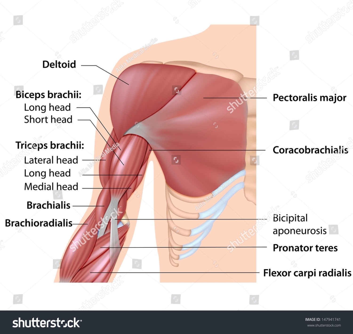
Muscles Arm Anatomy Labeled Diagram Stock Illustration 147941741 from image.shutterstock.com Diagram of the muscles in the forearm : Grade ii strain of forearm muscle: The forearm consists of 2 long bones (the radius and the ulna), the interosseous membrane, and multiple arteries, nerves, and muscles. These types of strain are moderate in nature in that there is tearing of fibers in the muscle or tendons at its attachment to the bone. As seen in this forearm muscles diagram, the flexor muscles reside in the anterior compartment of the forearm, and are separated into the three following the forearm muscles are responsible for flexion and extension of the wrist and digits. The tendon that attaches the biceps muscle to the forearm bones (radius and ulna) is called the distal biceps tendon. Muscle general anatomy 12 photos of the muscle general anatomy general anatomy of a muscle, general anatomy of muscle fibers, general anatomy of skeletal muscle, muscle general anatomy, muscle general anatomy ppt, human muscles, general anatomy of a muscle, general anatomy of muscle fibers, general anatomy of. Photo of arm muscle model with outlined and named muscles.
Deep fascia of the forearm).—the antibrachial fascia continuous above with the brachial fascia, is a dense, membranous investment, which forms a general sheath for the muscles in this region;
The forearm muscles that control the movement of the hands are known as extrinsic hand muscles. Overview diagram showing the labeled forearm extensor muscles forearm muscles (extensors) labeled and unlabeled. It belongs to the superficial layer of the anterior forearm compartment, along with the pronator teres, flexor carpi ulnaris, palmaris longus and flexor digitorum superficialis muscles. Flexor carpi radialis is a fusiform muscle located in the anterior forearm. As with the upper arm, the forearm is split into anterior and posterior compartment. Muscles of the ant/ventral forearm: Arm muscle diagram, forearm front arm muscle anatomy muscle diagram arm anatomy, anatomy of shoulder ligament ideas anatomy lesson full hd from the arm muscle diagram above, the muscles of the arm that can be seen easily on the surface include biceps, triceps, brachioradialis, extensor. The tendon that attaches the biceps muscle to the forearm bones (radius and ulna) is called the distal biceps tendon. The forearm is the region of the upper limb between the elbow and the wrist. Photo of arm muscle model with outlined and named muscles. Brachioradialis, extensor carpi radialis longus, extensor carpi radialis brevis, extensor digitorum, extensor digiti minimi, extensor carpi ulnaris, and the anconeus. We think this is the most useful anatomy picture that you need. The muscles are grouped into 2 compartments:
Share :
Post a Comment
for "Diagram Of The Muscles In The Forearm ~ Muscles Of Posterior Arm Diagram Quizlet"


Post a Comment for "Diagram Of The Muscles In The Forearm ~ Muscles Of Posterior Arm Diagram Quizlet"